How optical filters are used in positron emission tomography
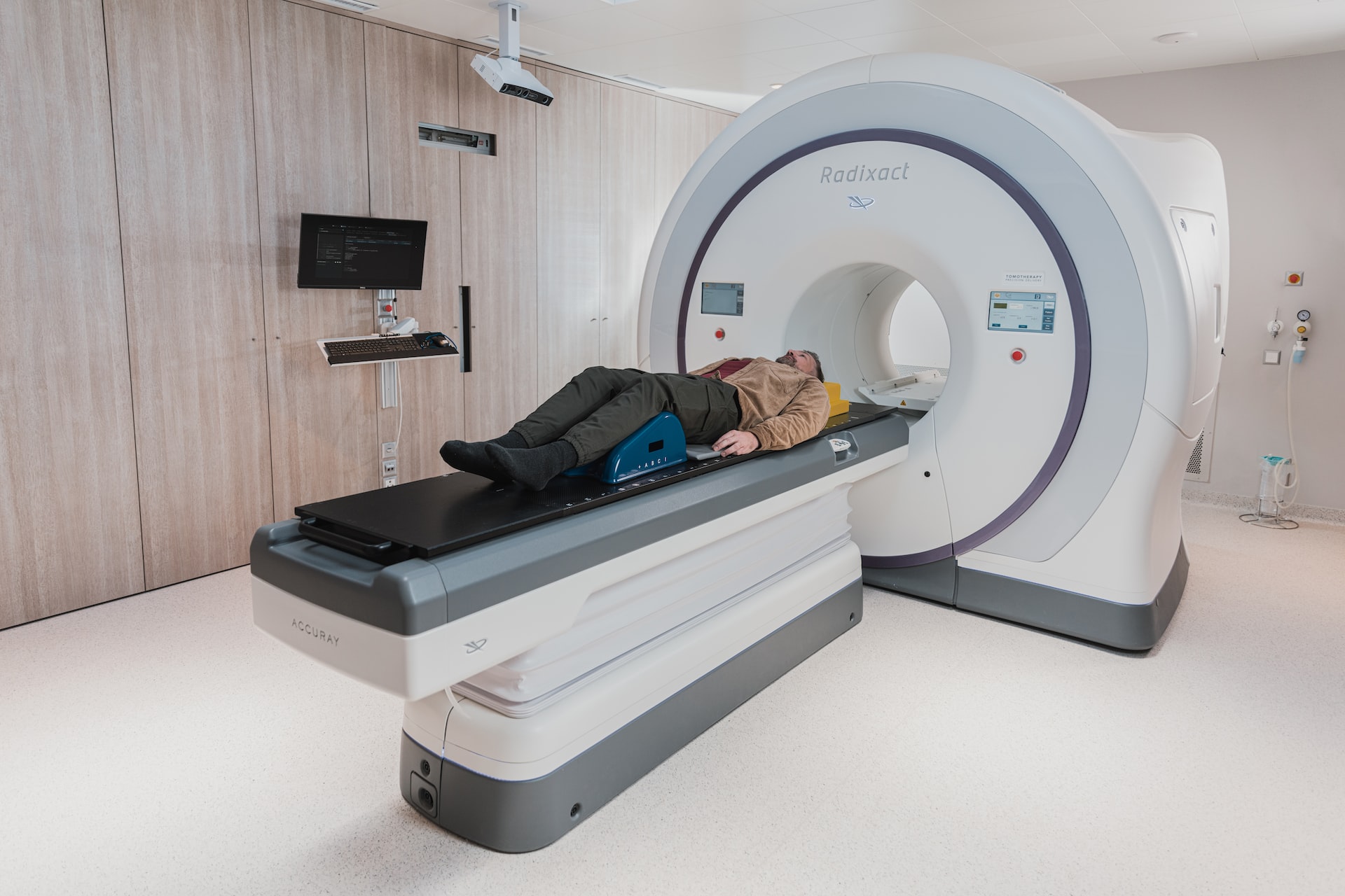
Positron emission tomography (PET) is one of a number of imaging techniques used in medical diagnostics. It uses a radiopharmaceutical agent to produce an image from inside the body, without using conventionally ‘invasive’ methods.
While the patient is exposed to a small amount of radiation – and should avoid pregnant women and very young babies for several hours after the procedure – PET is generally considered to be a safe way to obtain a 3D image from inside the body.
How does a PET scan work?
Briefly, a radioactive compound is injected into the body and metabolised by the patient’s cells, including cancer cells. For example, the compound F-18 fluorodeoxyglucose (FDG) is metabolised to FDG-6-phosphate.
Tumour cells cannot metabolise FDG-6-phosphate further, so it collects in cancerous tissue and can then be detected by scanning for the emission of positrons. These in turn are detected by looking for 511 keV photons emitted when a positron annihilates with an electron.
Creating the PET image
As the photons leave the body, they interact with the patient’s tissue via a process called Compton scattering. This interaction between light and matter is very similar to Rayleigh scattering, the process by which the sky appears blue in colour.
For approximately every 7 cm the beam travels through soft tissue in the body, its intensity halves (i.e. by 14 cm it will have one quarter of its original intensity, by 21 cm one eighth, and so on).
By detecting this beam from two opposing directions, the relative intensities can be used to reconstruct a single ‘voxel’ (a unit of volume, or a 3D pixel) in the overall PET image.
PET and other imaging techniques
PET can be used in combination with other medical imaging techniques, some of which are more familiar to the layperson:
CT (Computed Tomography, previously known as Computerised Axial Tomography)
MRI (Magnetic Resonance Imaging)
By combining PET with CT, MRI and other diagnostic imaging methods, more detailed data can be obtained, overcoming some of the limitations of using PET alone.
Optical filters for medical imaging
Optical filters play an important role in ensuring that the data obtained via medical imaging techniques is as accurate and detailed as possible.
Part of the reason for using imaging technology in medicine is the belief that computer-aided scans can identify details so fine that they would not be visible to the human eye. This is in addition to the ability to scan internal organs without the need to operate.
For this reason, the filters used in medical devices need to be of very high quality. Any defects can adversely affect the resolution of the data obtained during a scan, and could lead to important details being missed.
How optical filters help in medical scans
In a PET scan, a scintillation detector is used to measure the intensity of photons escaping the patient’s body.
The detector uses a crystal which, when encountered by a 511 keV photon, emits a burst of photons with much lower individual energy, placing them on (or near to) the visible light spectrum.
Highly detailed optical filters with purpose-built bandpass characteristics are used to ensure a ‘clean’ signal, such that the intensity of the visible light is proportional to the intensity of the 511 keV photon beam leaving the body.
Bespoke optical filters for medical imaging
Envin Scientific have the technology to design, build and – crucially – to accurately test the performance of optical filters in the 175 nm to 8 microns wavelength range, using Perkin Elmer spectrophotometers to verify the profile of the finished filter.
To find out more, or to ask us about bespoke optical filters for medical imaging of any type, please contact Envin Scientific today.